Glistening Ink-Cap (Coprinellus micaceus)
Glistening Inkcap (Coprinellus micaceus) is a common species of fungus in the family Psathyrellaceae with a cosmopolitan distribution.
The fruit bodies of the saprobe typically grow in clusters on or near rotting hardwood tree stumps or underground tree roots. Depending on their stage of development, the tawny-brown mushroom caps may range in shape from oval to bell-shaped to convex, and reach diameters up to 3 cm (1.2 in). The caps, marked with fine radial grooves that extend nearly to the center, rest atop whitish stems up to 10 cm (3.9 in) long. In young specimens, the entire cap surface is coated with a fine layer of reflective mica-like cells that provide the inspiration for both the mushroom's species name and the common names mica cap, shiny cap, and glistening inky cap. Although small and with thin flesh, the mushrooms are usually bountiful, as they typically grow in dense clusters. A few hours after collection, the gills will begin to slowly dissolve into a black, inky, spore-laden liquid—an enzymatic process called autodigestion or deliquescence. The fruit bodies are edible before the gills blacken and dissolve, and cooking will stop the autodigestion process.
The microscopic characteristics and cytogenetics of C. micaceus are well-known, and it has been used frequently as a model organism to study cell division and meiosis in Basidiomycetes. Chemical analysis of the fruit bodies has revealed the presence of antibacterial and enzyme-inhibiting compounds. Formerly known as Coprinus micaceus, the species was transferred to Coprinellus in 2001 as phylogenetic analyses provided the impetus for a reorganization of the many species formerly grouped together in the genus Coprinus. Based on external appearance, C. micaceus is virtually indistinguishable from C. truncorum, and it has been suggested that many reported collections of the former may be of the latter.
History and taxonomy[]
Coprinellus micaeus was illustrated in a woodcut by the 16th-century botanist Carolus Clusius in what is arguably the first published monograph on fungi, the 1601 Rariorum plantarum historia. Fungorum in Pannoniis observatorum brevis historia (History of rare plants. Brief history of fungi observed in Pannonia [Hungary]). Clusius erroneously believed the species to be poisonous, and classified it as a genus of Fungi perniciales(harmful fungi). The species was first described scientifically by French botanist Jean Baptiste François Pierre Bulliard in 1786 as Agaricus micaceus in his work Herbier de la France. In 1801, Christian Hendrik Persoongrouped together all of the gilled fungi that auto-digested (deliquesced) during spore discharge into the section Coprinus of the genus Agaricus. Elias Magnus Fries later raised Persoon's section Coprinus to genus rank in his Epicrisis Systematis Mycologici, and the species became known as Coprinus micaceus. It was thetype species of subsection Exannulati in section Micacei of the genus Coprinus, a grouping of related taxa with veils made of sphaerocysts (round swollen cells usually formed in clusters) exclusively or with thin-filamentous connective hyphae intermixed. Molecular studies published in the 1990s demonstrated that many of the coprinoid (Coprinus-like) mushrooms were in fact unrelated to each other. This culminated in a 2001 revision of the genus Coprinus, which was split into four genera; C. micaeus was transferred to Coprinellus.
Due partly to their ready availability and the ease with which they may be grown in the laboratory, C. micaceusand other coprinoid mushrooms were common subjects in cytological studies of the 19th and 20th centuries. The German botanist Johann Heinrich Friedrich Link reported his observations of the structure of the hymenium (the fertile spore-bearing surface) in 1809, but misinterpreted what he had seen. Link thought that microscopic structures known today as basidia were thecae, comparable in form to the asci of the Ascomycetes, and that each theca contained four series of spores. His inaccurate drawings of the hymenium of C. micaceus were copied in subsequent mycological publications by other authors, and it was not until microscopy had advanced that mycologists were able to determine the true nature of the basidia, when nearly three decades later in 1837 Joseph-Henri Léveilléand August Corda independently published correct descriptions of the structure of the hymenium. In 1924, A. H. Reginald Buller published a comprehensive description and analysis of the processes of spore production and release in the third volume of his Researches on Fungi.
The specific epithet micaceus is derived from the Latin word mica, for "crumb, grain of salt" and the suffix -aceus, "like, similar"; the modern application of "mica" to a very different substancecomes from the influence of micare, "glitter". The mushroom is commonly known as the "shiny cap", the "mica cap" or the "glistening inky cap", all in reference to the mealy particles found on the cap that glisten like mica.
Description[]
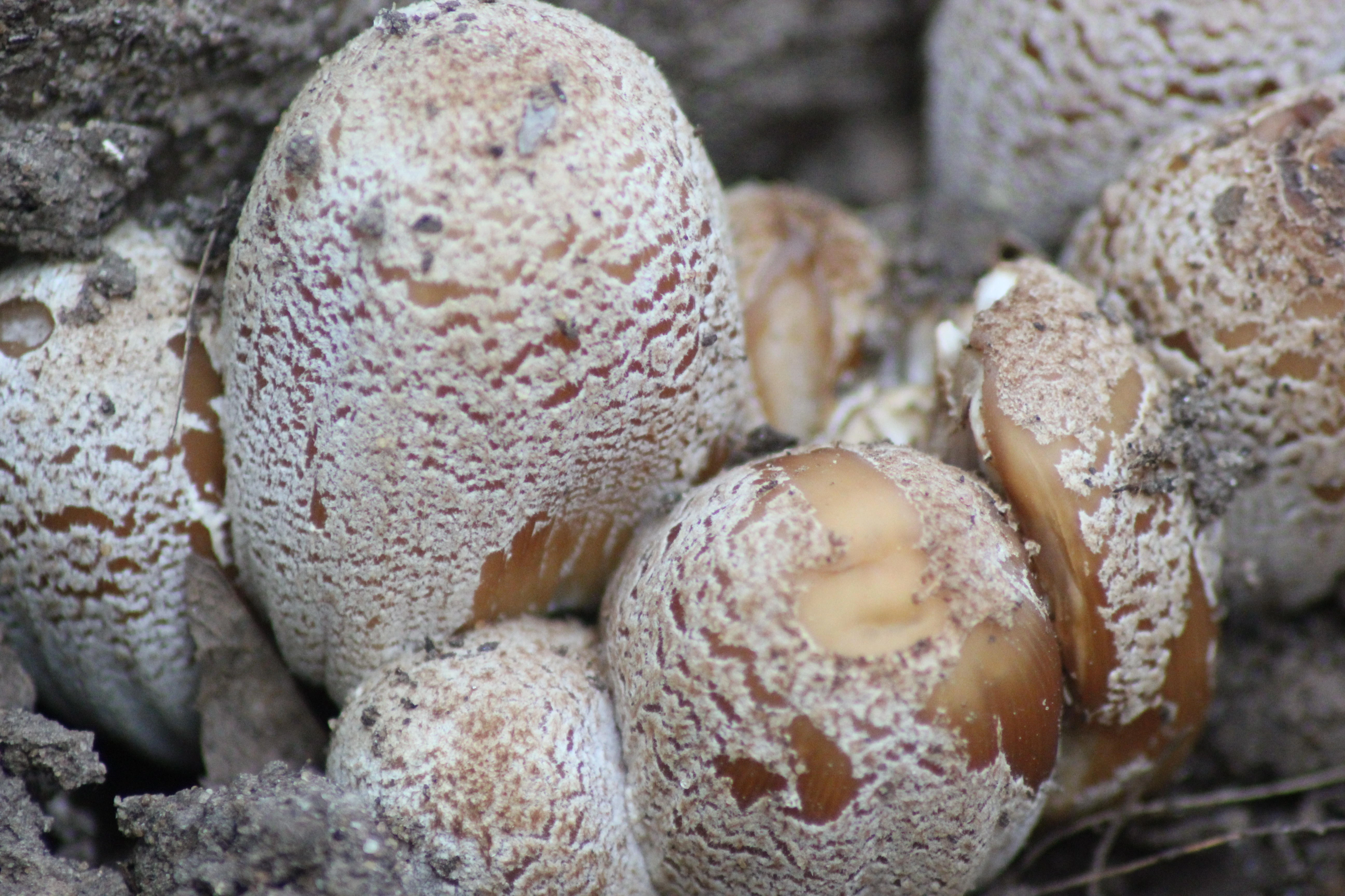
The cap is initially 1–2.5 cm (0.4–1.0 in) in diameter, oval to cylindrical, but expands to become campanulate (bell-shaped), sometimes with an umbo (a central nipple-like protrusion); finally it flattens somewhat, becoming convex. When expanded, the cap diameter reaches 0.8–3.0 cm (0.3–1.2 in) with the margin torn into rays and turned upwards slightly. The color is yellow-brown or tan often with a darker center, then pale yellow or buff from the margin inwards. The cap margin is prominently grooved almost all the way to the center; the grooves mark the positions of the longer gills on the underside of the cap. When young, the cap surface is covered with white or whitish shiny particles, remnants of the universal veil that covers immature specimens. The particles are loosely attached and easily washed away, so that older specimens are often smooth. Coprinellus micaceus is hygrophanous, meaning it assumes different colors depending on its state of hydration.
The gills are crowded together closely, and have an adnexed (narrow) attachment to the stem. Initially white, they change color to dark brown then eventually black as the spores mature. Expansion of the cap causes the gills to split open down their median planes, tearing the cap margin into rays. The process of spore discharge and autodigestion begin at the bottom of the gills before the upper parts of the gills have become completely blackened. The brittle stem is hollow, and measures 4–10 cm (1.6–3.9 in) long by 0.2–0.5 cm (0.1–0.2 in) thick and is roughly the same diameter throughout the length of the stem. It is generally white, but may discolor to pale dirty cream from the base up. The stem surface is at first velvety with a very fine whitish powder, but this eventually wears off, leaving it more or less smooth. Stems may have a rudimentary ring at the base, another universal veil remnant. The spore print is dark brown or black. The flesh is thin, fragile, white in the stem, and brownish in the cap. Its odor and taste are not distinctive. Individual fruit bodies take an average of five to seven days to fully mature.
Microscopic characteristics[]
The spores of C. micaceus are reddish-brown, with dimensions of 7–10 by 4.5–6 µm. Generally, they are lentiform (shaped like a biconvex lens), but viewed from the side they appear more almond-shaped or spindle-shaped, while in front view they appear oval or mitriform (roughly the shape of a miter—a peaked cap). Spores have a germ pore, a flattened area in the center of the spore surface through which a germ tube may emerge. The spore-bearing cells (the basidia) are four-spored, club-shaped, and measure 10–15 by 4–7 µm. Studies have shown that the basidia develop in four discrete generations. The first generation basidia are the most protuberant, and extend out the greatest distance from the surface of the hymenium. Subsequent generations of basidia have shorter and less protuberant bodies. When a living gill is viewed with a microscope, the four sets of basidia can be seen distinctly. Arthur Buller coined the term inaequihymeniiferous to describe this mode of hymenial development. The purpose of the staggered basidia sizes is to facilitate the release of spores from the hymenium. There are four zones of spore discharge that correspond to the four sets of basidia, and basidia that have released all of their spores quickly begin to autodigest. The staggered setup minimizes the chance of spores colliding with neighboring basidia during release.
Cystidia that are located along the edge of the cap (called cheilocystidia) are spherical, and 30–120 by 20–74 µm. The facial cystidia (called pleurocystidia) are club-shaped or elongated ellipses, up to 130–155 µm in length. The pleurocystidia protrude from the face of the gill and act as guards, preventing adjacent gills from touching each other, and also ensuring that the basidia and spores have sufficient room for development. C. micaceus may also have scattered caulocystidia (cystidia on the stem) that are 60–100 by 5–10 µm, but their presence is variable and cannot reliably be used for identification. Both De Bary and Buller, in their investigations into the structure of the cystidia, concluded that there is a central mass of cytoplasm formed where numerous thin plates of cytoplasm meet at the center of the cell. De Bary believed that the plates were filamentous branching processes,[ but Buller thought that they were formed in a process similar to the walls of foam bubbles, and that the central mass was able to slowly change form and position by altering the relative volumes of the vacuoles enclosed by the numerous thin cytoplasmic walls. In older cells, the cytoplasm may be limited to the periphery of the cell, with one huge vacuole occupying the cell center.
The globular cells that make up the mica-resembling scales on the cap are colorless, smooth-walled, and range in size from about 25–65 µm, although most are between 40–50 µm. Buller explained the "glitter" of these cells as follows: "The sparkling of the meal-cells, as well as of the cystidia on the edges and faces of the gills, is simply due to light which strikes them from without and is refracted and reflected to the eye in the same manner as from the minute drops of water one so often sees at the tips of grass leaves on English lawns early in the morning after a dewy night."
Edibility[]
Coprinellus micaceus is an edible species, and cooking inactivates the enzymes that cause autodigestion or deliquescence—a process that can begin as soon as one hour after collection. It is considered ideal for omelettes, and as a flavor for sauces, although it is "a very delicate species easily spoiled by overcooking". The fungus also appeals to fruit flies of the genus Drosophila, who frequently use the fruit bodies as hosts for larvae production.
A study of the mineral contents of various edible mushrooms found that C. micaceus contained the highest concentration of potassium in the 34 species tested, close to half a gram of potassium per kilogram of mushroom. Because the species can bioaccumulate detrimental heavy metals like lead and cadmium, it has been advised to restrict consumption of specimens collected from roadsides or other collection sites that may be exposed to or contain pollutants.
Ecology, habitat and distribution[]
Coprinellus micaceus is a saprobic species, deriving nutrients from dead and decomposing organic matter, and grows in and around stumps or logs of broad-leaved trees or attached to buried wood. It prefers feeding on bark, particularly the secondary phloem, rather than the wood. In the scheme of the succession of fungal species involved in the decomposition of wood, C. micaceus is a late stage colonizer, and prefers to feed on wood that has already decomposed sufficiently to have reached "a friable softened consistency". A 2010 study suggests that the fungus can also live as an endophyte, inhabiting the woody tissue of healthy trees without causing disease symptoms. The fungus is also associated with disturbed or developed ground, such as the sides of roads and paths, gardens, building sites and the edges of parking lots; it has also been noted for growing indoors on rotting wood in humid environments. In one instance it was discovered about 120 m (400 ft) underground in an abandoned coal mine, growing on wooden gangways and props used to support the roof.
Fruit bodies are commonly found growing in dense clusters, but can also be found growing singly or in small clumps, especially in forested areas. In North America, C. micaceus is one of the first edible mushrooms to appear in the spring, and fruits from May to September. In Europe, it fruits from May to December. Although it can grow at any time of the year, it is more prevalent during the spring and fall, coinciding with the higher humidity resulting from spring and autumn rains. The species is known for reappearing with successive fruitings at the same location. In one case, a total of 38 lb (17.2 kg) of fresh mushrooms were collected from one elm stump in 10 successive crops over a spring and summer.
Coprinellus micaceus has a cosmopolitan distribution, and has been collected in northern Africa, South Africa, Europe (including Turkey), North America (as far north as Alaska), the Hawaiian islands, South America, India, Australia, New Zealand, and Japan. Phylogenetic analysis of rDNA sequences from specimens collected in southeastern Asia and Hawaii show that the Hawaiian species form a distinct clade with little genetic diversity compared to Asian populations; this suggests that the Hawaiian populations have been introduced relatively recently and have not had much time to develop genetic variation. One study suggests that in South Africa, where C. micaceus is rare, it has been frequently confused with the similar-appearing C. truncorum, a more common species in that region. A similar inference has been raised about North American species.